Optical fibres will help peek into the deepest parts of the brain. A doctoral student from FME is involved in the research
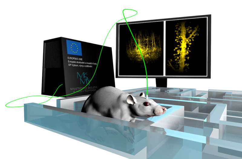
To look through an optical fibre thinner than a human hair inside the human brain. This is the ambition of the scientific team, which includes the doctoral student Miroslav Stibůrek from the FME. So far, experts are testing the so-called holographic micro-endoscopy on mice, but in the future, they would like this tissue-friendly and detailed imaging method to be accessible to human patients as well.
Many people are familiar with endoscopic examinations, such as of the oesophagus or intestines, where a catheter equipped with a camera and other instruments is inserted into the body. These examinations are not particularly pleasant. In the future, the use of smaller probes made of optical fibres could make it easier for patients. Experts from the Institute of Scientific Instruments (ISI) of the Czech Academy of Sciences, whose team includes the doctoral student from the Faculty of Mechanical Engineering at BUT and a graduate of FEEC, Miroslav Stibůrek, are working on the development of these fibres. This year, Stibůrek received an internal BUT grant for young scientists, known as KInG, for research on optical fibres for the human brain imaging.
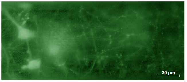
"People mainly associate fibre optics with fast internet. However, we are able to use a single optical fibre, as thin as a human hair, to image structures inside living tissues. This is at submicron resolution, meaning we can see down to the level of cell organelles," Stibůrek explains.
Compared to current methods of brain examination, optical fibres offer a much more detailed view into the tissue's depth, which devices like CT or tomography cannot achieve. "Today it is always a quid pro quo: if we can image tissues at any depth, we lose the ability to see details; the resolution of devices is usually in millimetres. With light microscopic methods, we achieve great resolution but cannot penetrate depth. Holographic micro-endoscopy allows us to reach any depth and achieve a resolution comparable to light microscopy," Stibůrek adds.

He is part of the team led by Professor Tomáš Čižmár at the Academy of Sciences. This year, he received a half-million grant from BUT for young doctoral students, which will allow him to spend a year researching the process of preparing optical fibre. "Cutting the fibre and inserting it into the brain is not enough for us. We deposit various layers of metals and polymers, with a thicknesses of several units to hundreds of nanometres. I want to focus on this process of preparation in my work this year," Stibůrek explains, believing that in the future, the research could also turn into a startup that could bring this promising technology to the market and thus to hospitals.

(ivu)
| Published | |
|---|---|
| Link | https://www.vut.cz/en/alumni/f38103/d225453 |
Responsibility: Mgr. Marta Vaňková