FIT BUT researchers help develop revolutionary 3D scanner for better diagnosis of vascular diseases and arthritis
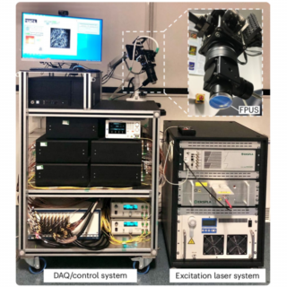
A research team led by Associate Professor Jaroš from the Faculty of Information Technology at Brno University of Technology (FIT VUT), in collaboration with University College London (UCL), contributed to the development of groundbreaking diagnostic technology – an optical 3D photoacoustic scanner. This innovative method opens new possibilities for non-invasive diagnostics of vascular diseases, inflammatory skin conditions, and rheumatoid arthritis. The technology not only shortens examination time but also ensures significantly more accurate results. The research findings were published in the prestigious journal Nature Biomedical Engineering, a leader in applied medical informatics.

How does the technology work?

The optical 3D photoacoustic scanner resolves this issue by optimizing software and using advanced data processing methods. Since 2014, the development of this technology has been driven by a team of Brno researchers from FIT VUT under the leadership of Associate Professor Jiří Jaroš. Their efforts resulted in a dramatic acceleration of computational processes associated with photoacoustic tomography. The new scanner can produce a 3D image within seconds, eliminating sensitivity to patient movement and enabling practical clinical applications.
“The technology we have been working on since 2014 is finally finding real-world applications in medicine. By improving speed and accuracy, we have overcome the limitations that previously prevented this method from being used in clinical practice,” said Associate Professor Jiří Jaroš from FIT VUT.
Practical Benefits for Medicine

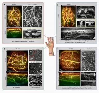
One of the key benefits of the scanner is its ability to provide detailed visualization of vascular structures in the early stages of disease. For diabetic patients, the scanner can detect changes in microcirculation that often precede serious complications, such as peripheral vascular disease. In the case of rheumatoid arthritis, it enables monitoring of new blood vessel formation in affected areas, providing doctors with valuable information to assess inflammatory activity and optimize treatment.
In dermatology and oncology, the scanner offers entirely new possibilities for monitoring vascular changes, for example, in skin inflammations or tumor growth. By capturing dynamic changes in the vascular system, it significantly surpasses the limitations of current ultrasound methods, enabling more precise diagnostics without unnecessary delays.
Path to a Breakthrough Technology
The development of the scanner is a prime example of international collaboration and interdisciplinary approaches. The project's initial steps involved two bachelor theses at FIT VUT, focused on accelerating computational processes, followed by transitioning from the programming language Matlab to highly optimized C++ code that fully utilizes multi-core processors. The code was further accelerated using a graphics card, leading to a twentyfold increase in computational speed. The team led by Associate Professor Jaroš worked on software optimization, which is now integrated into the LabVIEW environment and further developed in collaboration with University College London (UCL). “Our collaboration with UCL was key. Together, we optimized not only software computations but also the scanner's hardware itself. This allowed us to minimize device delays,” explained Associate Professor Jaroš.
Publication in *Nature Biomedical Engineering*
The technology's results were published in the article A fast all-optical 3D photoacoustic scanner for clinical vascular imaging. The publication not only confirms the technological superiority of the device but also demonstrates its benefits in imaging vascular structures in patients with diabetes, skin inflammation, and rheumatoid arthritis.
The future of diagnostics
A new scanner paves the way for a revolution in medical diagnostics. "In the future, the technology may find applications in real-time monitoring of dynamic vascular changes or in the study of parameters related to blood flow in patients with cardiovascular disease. Its use thus promises better and faster care for patients and new possibilities for medical research," said Associate Professor Jaroš in conclusion.
The text was published as a press release.
| Author | Mgr. Petra Horná |
|---|---|
| Published | |
| Link | https://www.vut.cz/en/but/f19528/d275130 |